The pursuit of a flawless smile has transitioned from a luxury reserved for the elite to a sophisticated branch of restorative science. Dental veneers—ultra-thin shells of material bonded to the front of teeth—represent a pinnacle of bio-engineering. While early iterations often resulted in an unnaturally opaque “piano key” appearance, modern cosmetic dentistry leverages advanced material science and light-refraction principles to mimic the organic complexity of human enamel.
The Biomimetic Evolution of Materials
The core of a natural-looking veneer lies in its ability to replicate the optical properties of a tooth. Natural enamel is translucent, meaning it allows light to penetrate the surface and reflect off the underlying dentin. Modern lithium disilicate and leucite-reinforced glass-ceramics are engineered with varying levels of opacity and translucency to recreate this depth.
Unlike older feldspathic porcelains, which were prone to brittleness, these high-tech ceramics undergo a crystallization process that increases their structural integrity. This allows dentists to create veneers as thin as 0.3mm, often requiring little to no removal of the healthy tooth structure. By maintaining the majority of the natural enamel, the bond strength is significantly higher, as the adhesive resins interface more effectively with enamel than with the deeper dentin layer.
Adhesion and Chemical Bonding
The “science” of the smile is held together by complex chemistry. The bonding process involves a multi-step conditioning sequence. First, the internal surface of the ceramic is etched with hydrofluoric acid to create microscopic porosities. Simultaneously, the tooth is treated with a phosphoric acid gel.
A silane coupling agent is then applied to the veneer, acting as a bridge between the inorganic ceramic and the organic resin cement. This creates a monolithic bond that, once light-cured, essentially fuses the veneer to the tooth. This chemical integration is what allows a thin sliver of porcelain to withstand the immense mechanical forces of mastication, which can exceed 700 Newtons in the molar region.
Digital Shade Matching and Layering
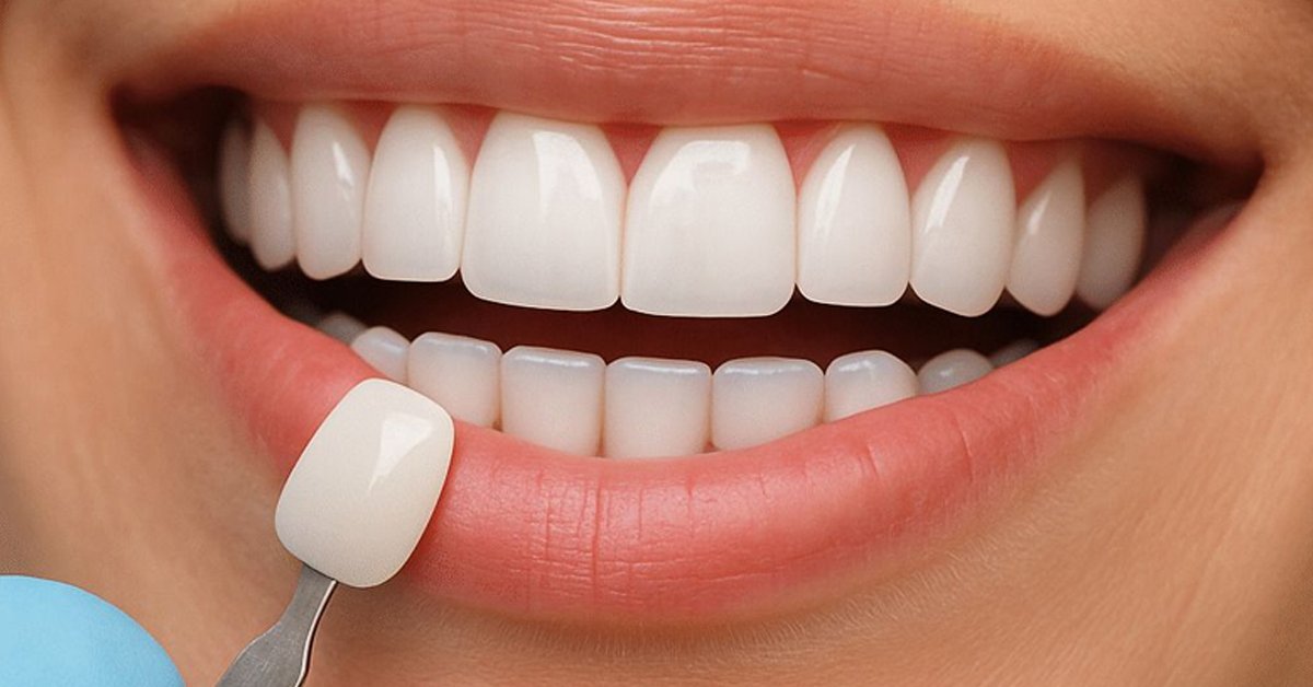
One of the most significant hurdles in cosmetic dentistry has been “metamerism”—the phenomenon where a veneer looks perfect under dental office LED lights but appears grey or unnaturally white in natural sunlight. To combat this, clinicians now use digital spectrophotometers to measure the exact hue, value, and chroma of the surrounding teeth.
In the laboratory, technicians use a multi-layered approach. Instead of a single uniform block of colour, they build the veneer using internal staining techniques. By placing warmer, more opaque tints at the cervical (gum) margin and increasing translucency toward the incisal (biting) edge, the final restoration captures the “halo effect” seen in youthful, natural teeth.
The Rise of Composite Innovations
While porcelain remains the gold standard for longevity, the industry has seen a massive shift toward resin-based solutions. We are currently witnessing the rise of composite veneers in digital cosmetic dentistry as a highly versatile alternative. Modern nanofilled composites contain sub-micron glass particles that allow the material to be polished to a high lustre, rivaling the light reflection of ceramic.
This technique is particularly popular for “chairside” transformations. Because the resin is applied incrementally, the dentist can sculpt the anatomy of the tooth in real-time, adjusting the contours to complement the patient’s lip line and facial symmetry. The science here focuses on polymerisation shrinkage control, ensuring the material remains stable and stain-resistant over years of use.
Periodontal Integration and Biological Width
A beautiful veneer is a failure if it compromises the health of the gingiva. The science of “biological width” dictates exactly where the veneer margin should sit. If a restoration is placed too deep beneath the gum line, it triggers a chronic inflammatory response, leading to redness and recession.
Modern techniques utilize retraction cords and digital Scanners to ensure the margin is “equigingival” or slightly “supragingival.” This precision prevents the accumulation of biofilm and ensures that the transition between the synthetic material and the natural gum tissue is seamless. When executed correctly, the gingival tissue remains a healthy coral pink, framing the new smile without any signs of “dark lines” at the margin.
The Precision of Digital Workflows
The marriage of artistry and engineering is best seen in the transition from physical impressions to digital “twins.” Traditional putty impressions were prone to dimensional instability, often shrinking or warping by fractions of a millimetre—enough to cause a misfit.
Today’s CAD/CAM (Computer-Aided Design and Computer-Aided Manufacturing) systems allow for a level of accuracy that was previously impossible. By combining the precision of technology with biological principles, practitioners can now mill veneers from solid blocks of ceramic with marginal gaps measured in microns. This “digital smile design” allows patients to “test drive” their smile via a 3D-printed mock-up before a single tooth is touched, ensuring the final result is as functional as it is aesthetic.
READ ALSO: Dental Health Innovations: Exploring the Future of Implants